
Introduction:
The major characteristic that differentiates between living organisms and non-living is the
Locomotion or movement. This feature of the movement is very crucial for living organisms as they need to change themselves according to the environment which helps them to get sustain themselves in this world.
Every organism has its special system for locomotion and movement such as cilia, and flagella ( rudimentary structures) or dynamic systems like wings or feet. According to the scientist’s theory, locomotion and movement played a significant role in the process of evolution of humans – from being quadrupedal to bipedal and increasing the brain’s volume.
Types of Joints:
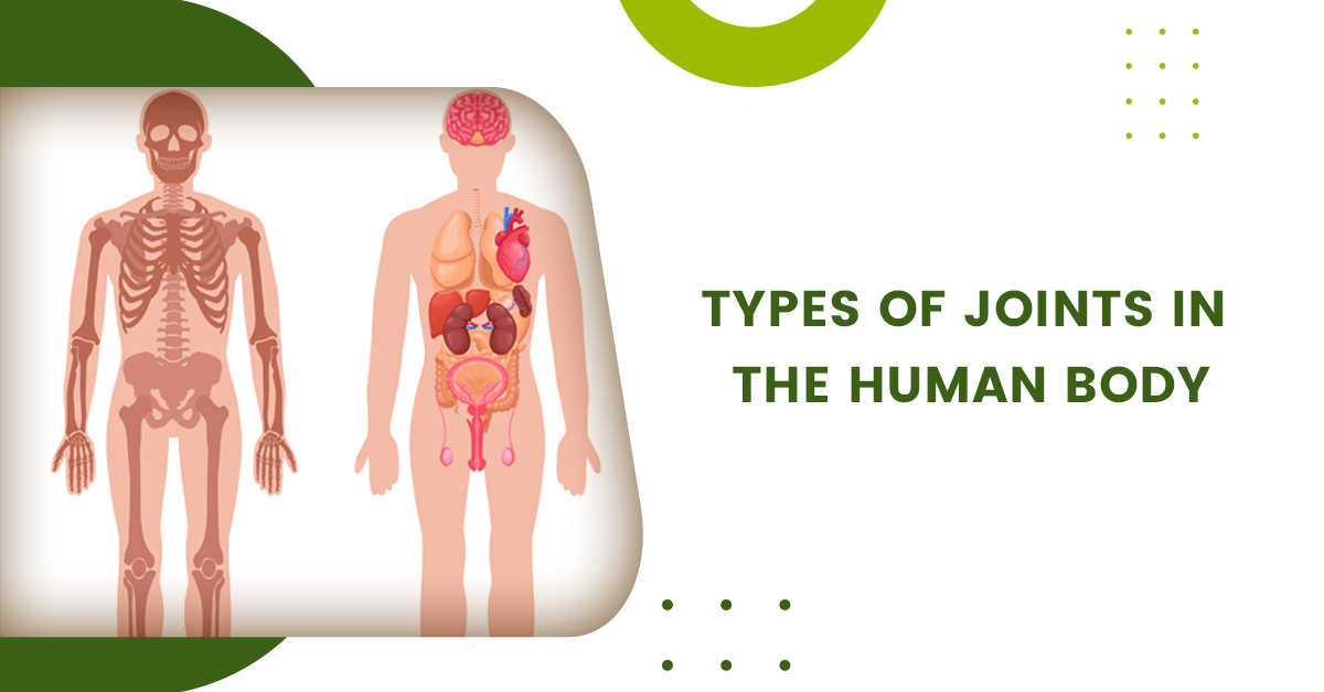
A joint is a location at which two bones meet. An articulation or articular surface is a joint connecting two bones in the skeletal system. Joints provide the ability to move our body parts.
Joints can be classed histologically based on the predominant kind of connective tissue or functionally based on the range of motion allowed. Fibrous, cartilaginous, and synovial joints are the three types of joints in the body based on structure.
Based on functionality there are three types of joints synarthrosis (immovable), amphiarthrosis (slightly moveable), and diarthrosis (freely moveable). There is a correlation between the two classification schemes: synarthroses have fibrous tissues, amphiarthroses have cartilaginous tissues, and diarthrosis has synovial tissues.
Fibrous Joint-
Fibrous Types of Jointsare the joints where fibrous tissue comprises primarily collagen that connects bones which are fixed in nature. So we can say that usually fibrous joints are synarthroses (immoveable) and have no joint cavity. These joints are further subdivided into sutures, gomphosis, and syndesmoses.
- Sutures are cranial joints that are immovable. The connective tissue connecting the plate-like bones of the skull, known as fontanelles, makes them slightly movable after birth. This early flexibility enables the infant’s head to pass through the birth canal and allows the brain to expand after birth. Sharpey’s fibres, a thin layer of fibrous connective tissue that sutures the bone plates together, replace the fontanelles as the skull grows larger. The two adjacent plates combine to form one bone as cranial sutures ossify; this fusion is known as synostosis.
- The mandible and maxillae have immovable gomphosis that connects the teeth to their sockets. The fibrous tissue that links the tooth to the socket is called the periodontal ligament.
- Syndesmoses are joints that can move slightly (amphiarthroses). Syndesmosis joints are held together by an interosseous membrane. The middle tibiofibular joint, for example, is formed by the tibia connecting to the fibula, and the middle radio-ulnar joint is formed by the ulna connecting to the radius.
Cartilaginous Joint-
Hyaline cartilage or fibrocartilage connects the bones in cartilaginous joints. The joints are categorized as primary or secondary cartilaginous joints depending on the kind of cartilage involved.
- Synchondroses are primary cartilaginous joints that exclusively involve hyaline cartilage. Amphiarthroses are stationary joints that are slightly movable (synarthroses). A good example is a joint between the epiphysis and diaphysis of growing long bones.
- Hyaline or fibrocartilage may be involved in the secondary cartilaginous joint, commonly known as a symphysis. These joints move around a little (amphiarthroses). A pubic symphysis is a classic example.
Synovial Joint-
Synovial joints are the main functioning joints of the body and are freely movable (diarthrosis). The synovial joint is defined by its joint cavity. The articular capsule, a fibrous connective tissue linked to each participant’s bone slightly beyond its articulating surface, surrounds the hollow.
Located between the capsule and the joint cavity, synovial membranes (synovium) secrete synovial fluid. Articular cartilage is made up of hyaline cartilage that covers the whole articulating surface of each bone. Articular cartilage is attached to the synovium by ligaments. Some synovial joints have fibrocartilage between the articulating bones, such as menisci.
The types of movements that synovial joints allow are frequently classified further. Hinge (elbow), saddle (carpometacarpal joint), planar (acromioclavicular joint), pivot (atlantoaxial joint), condyloid (metacarpophalangeal joint), and ball and socket ( (hip joint) are the six classes.
1. Ball and Socket Joints
A bone is hooked into the hollow space of another bone in this position. Rotatory movement is aided by this joint. The shoulders are a type of ball and socket joint.
2. Pivotal Joints
This sort of joint occurs when one bone has tapped into the other, preventing full rotation. This joint helps you move sideways and back and forth. A crucial joint in the neck, for example.
3. Hinge Joints
In the same way that a door hinge can only move back and forth, hinge joints do the same. These joints include knees, elbows, and ankles.
4. Saddle Joints
The saddle joint is a biaxial joint that allows movement in two planes: flexion and extension, as well as abduction and adduction. The thumb, for example, is the only bone in the human body with a saddle joint.
5. Condyloid Joints
Condyloid joints have two axes and can move up and down as well as side to side. The base of the index finger, carpals of the wrist, elbow, and wrist joints all have condyloid joints. A condylar or ellipsoid joint is another name for this joint.
6. Gliding Joints
A common type of synovial joint is the gliding joint. It is also referred to as the plane or planar joint. Located between rounded or flat bones, this joint allows for free movement. This joint is mostly seen in areas where two bones contact and glide on each other in any direction. Gliding joints include the lower leg and ankle joint, as well as the forearm and wrist joint.
Anatomy of Joints-
The points where two or more bones touch are called joints. The bones can move due to the mobility of most joints. The following elements make up joints:
- The surface of a bone at a joint is covered with this sort of tissue. Cartilage serves to reduce friction during joint movement.
- The membrane of synovium The synovial membrane is a tissue that lines the joint and closes it into a joint capsule. To lubricate the joint, the synovial membrane secretes a clear, sticky fluid called synovial fluid.
- The joint is surrounded by strong ligaments (stiff, elastic bands of connective tissue) that provide support and limit movement. Ligaments join bones together.
- Tendons (a tough connective tissue) on both sides of a joint link to muscles that control the joint’s movement. Tendons form fibrous bands connecting muscles with bones.
- Bursas are fluid-filled sacs that sit between bones, ligaments, or other adjacent structures. They aid in reducing joint friction.
- Synovial fluid is a type of synovial fluid. The synovial membrane secretes a transparent, sticky fluid.
- In the knees and other joints, this is a curved piece of cartilage.
You also Can Read This – Body Aches – Causes, Symptoms, Treatment, Prevention